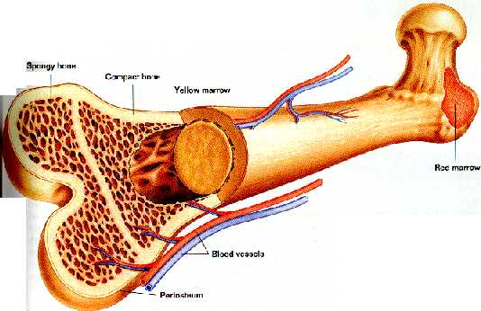
Bone marrow consists of two types, red(vascular) or yellow (fatty) bone marrow. Red bone marrow forms all blood cells, except lymphocytes, in humans. It also destructs old red blood cells. Yellow bone marrow stores fat. It can be converted into red bone marrow under certain conditions, like severe blood loss. All human bone marrow is red until about the age of seven because the need for new blood formation is high. Later, fat tissue slowly replaces the red bone marrow. Red bone marrow is found in adults only in the vertebrae, hips, breastbone, ribs, skull, and the ends of long bones. Yellow bone marrow fills the cavities of these bones.
Cancellous bone is very spongy in structure and appearance. It is made of trabeculae, which are rigid plates. They also have osteocytes with incomplete Haversian systems, so the osteocytes are nourished by a network of canaliculi.
Compact bone is a dense, ivory-like substance that surrounds the cancellous bone. It contains a series of ring deposits of mineral salts and collagen fibers, called lamellae. The lamellae are interlaced with the Haversian canals.
The periosteum is a sheath of tissue that covers the outer bone surfaces. During growth, it contains osteoblasts, and it contains connective tissue cells that can become osteoblasts that form bones in reaction to stress or injury. The periosteum is composed of two layers. One is an outer layer that provides a shield and a point of attachment for ligaments and tendons. The inner layer is called the endosteum, which is a thin membrane that contains bone forming and blood forming cells. It is also needed for bone remodeling.
Cartilage is a dense, connective tissue that covers the ends of bones. It is made of collagen fibers embedded within a substance which resembles plastic. This structure allows cartilage to be very strong, so it can support weight, while still being extremely flexible.
The first type of cartilage is hyaline cartilage which is the most common and it forms the embryonic skeleton. It exists in adults at the ends of bones in free moving joints, at the ends of ribs, and in the nose, trachea, larynx, and bronchi. The second type, fibrocartilage, is very tough, and found in disks which exist in side the vertebrae. The last type is yellow, or elastic, cartilage. It is the most flexible cartilage because it contains elastic fibers in addition to the collagen fibers. It forms the external ear, the auditory tube of the middle ear, and the epiglottis.
Another function of cartilage is to form a model for the growth of the bony skeleton. In the embryo, the fibrous part of the cartilage calcifies, and is replaced by chondrocytes, the cells that form cartilage, or osteocytes. In conclusion, cartilage is vital to the functions performed by the skeletal system.
There are two types of connective tissues. Ligaments are tough fibrous bands of connective tissue that support internal organs and hold bones together at the joints. A ligament is composed of dense fibrous bundles of collagenous fibers and spindle-shaped cells known as fibroblasts. (They are long, flat, spindle-shaped connective-tissue cells. Fibroblasts are vital in the production of collagen.) There are two major types of ligaments: white ligaments and yellow ligaments. White ligaments are rich in collagenous fibers. Yellow ligaments are rich in elastic fibers which allow movement.
At the joints, ligaments form a capsule-shaped sac that encloses bone ends and a lubricating membrane, the synovial membrane. Other ligaments fasten around or across bone ends in bands or act as a tie pieces between bones, such as in the ribs and forearm, restricting inappropriate movement. Tendons are the other type of tissue, that connects bones to muscles.


